
1. Case Presentation
A 15-year-old postmenarchal girl presented to the Emergency Department in June 2025 with sudden severe right lower-quadrant pain and nausea. Before this episode, she reported predominantly painless menses lasting 4–5 days and denied chronic pelvic pain, significant dysmenorrhea, or similar prior episodes. She had no other past medical or surgical history.
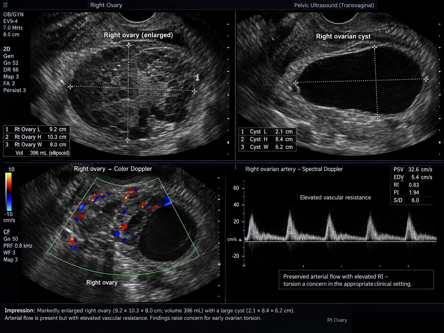
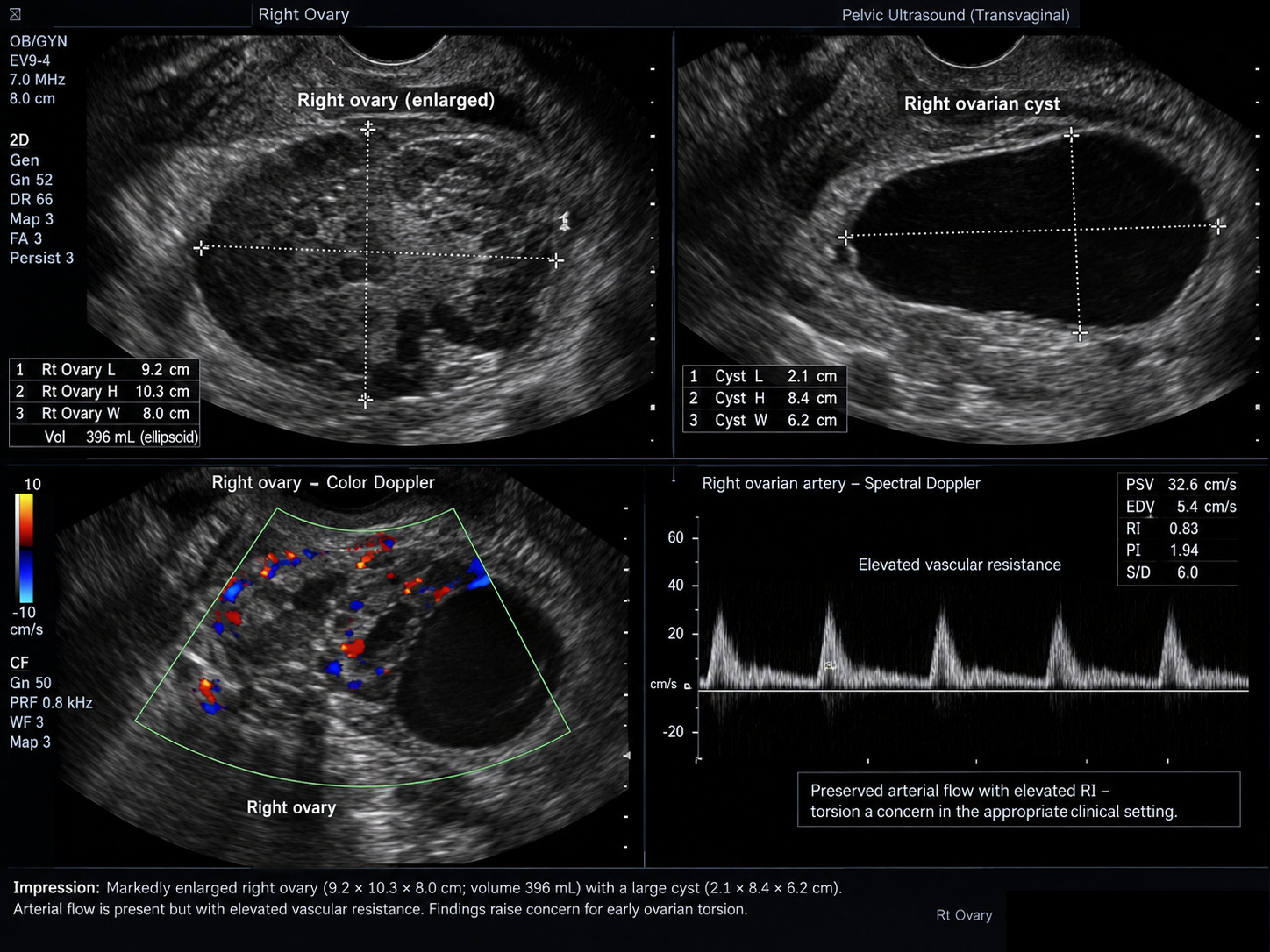
Pelvic ultrasonography reported a markedly enlarged right ovary measuring 9.2 × 10.3 × 8.0 cm (estimated volume, 396 mL) and an apparent ovarian cyst measuring 2.1 × 8.4 × 6.2 cm. Doppler flow was detectable but showed increased vascular resistance. A small amount of cul-de-sac free fluid was noted. The uterus and left ovary had no reported significant abnormality. Despite detectable flow, the clinical and sonographic findings raised concern for torsion (Figure 1).
Urgent laparoscopy confirmed right ovarian torsion. The right ovary was detorsed and preserved, with a satisfactory appearance after detorsion. The large cystic lesion, initially thought to be ovarian on ultrasound, was identified intraoperatively as originating in the right fallopian tube/peritubal region. The cyst was excised; no additional gross lesions suggestive of endometriosis were seen. The specimen was submitted as a right peritubal cyst. Final histopathologic examination showed endometriosis with vascular congestion and no malignancy.
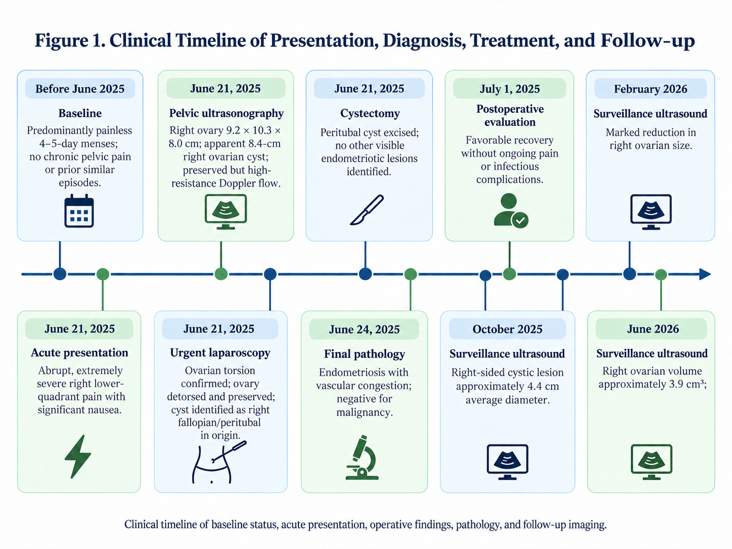
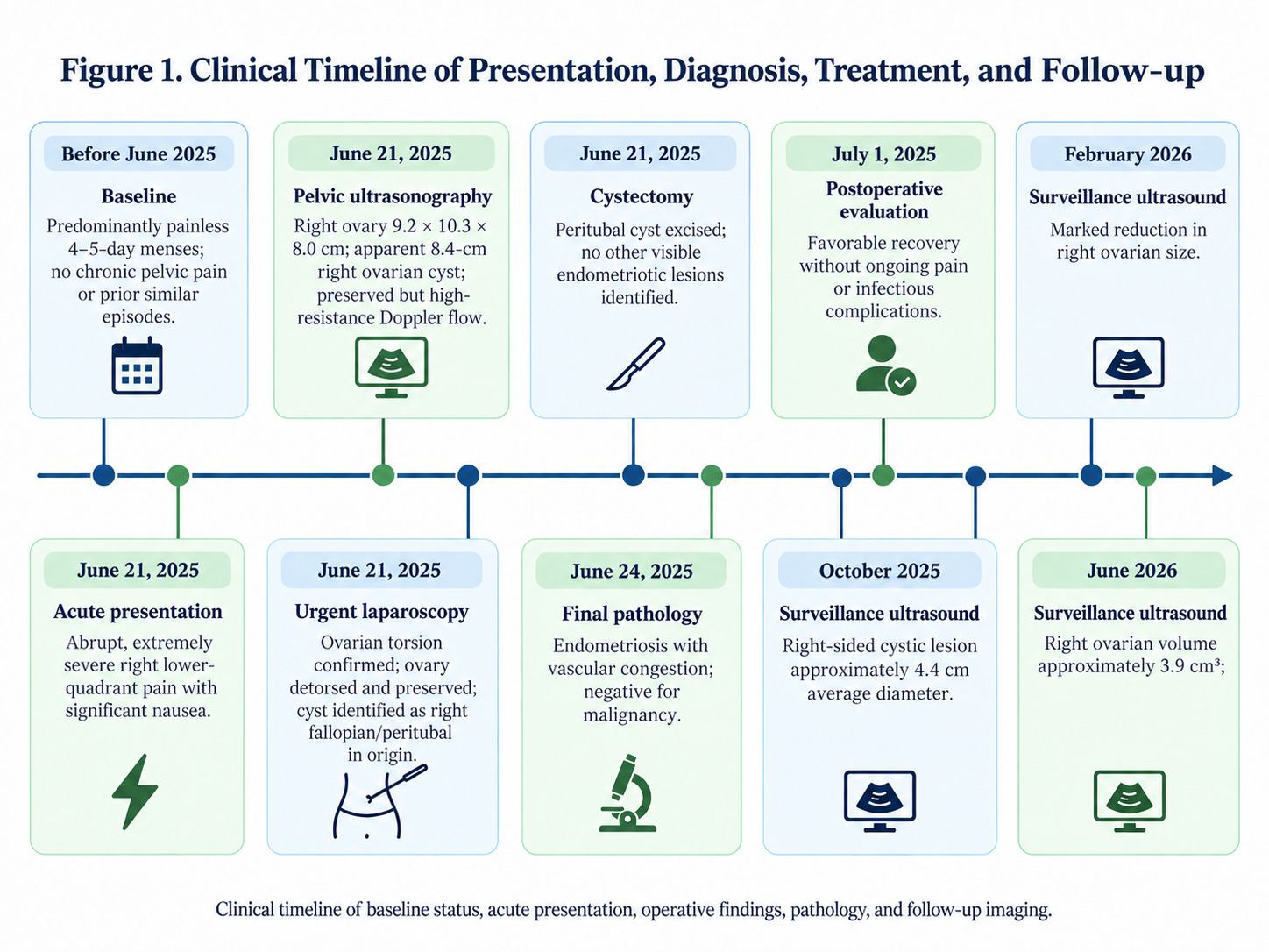
At approximately 2-week follow-up, the patient reported no ongoing abdominal pain, nausea, vomiting, or fever; her laparoscopic incisions were healing appropriately. Pathology was discussed with the patient and her mother, and gynecologic follow-up was arranged. In October 2025, ultrasound documented a right ovarian cyst measuring 49.1 × 37.7 × 44.5 mm. The February and June 2026 imaging records documented right ovarian volumes of approximately 9.2 and 3.9 cm³, respectively; however, it was unclear whether the October cyst had resolved or represented a new or persistent lesion. The chronology is summarized in Figure 2. This case report was prepared in accordance with the Consensus-based Clinical Case Reporting Guideline Development (CARE) guidelines.1
2. Discussion
Adolescent endometriosis is commonly considered in patients with dysmenorrhea or chronic pelvic pain.2,3 In a systematic review, endometriosis was identified in 648 of 1,011 (64%) adolescents who underwent laparoscopy for pelvic pain; that figure reflects a selected symptomatic group, not the prevalence in all adolescents.3 The current case illustrates a different diagnostic pathway: endometriosis was found histologically in a peritubal cyst removed during an acute surgical emergency, although the patient reported no preceding menstrual or chronic pelvic symptoms. The lack of additional visible implants does not exclude microscopic disease elsewhere.
Acute unilateral pain with nausea is typical of adnexal torsion. The presence of Doppler flow does not exclude torsion, and prompt laparoscopy with detorsion and adnexal preservation is recommended when the clinical suspicion is high.4–6 The surgical findings in this patient confirmed torsion despite preserved flow on ultrasound. In addition, the lesion interpreted preoperatively as ovarian was found to be peritubal. Large adnexal lesions can obscure their anatomic origin on ultrasonography, and operative inspection may clarify the relationship of a cyst to the ovary and fallopian tube.7,8
The pathologic finding supports endometriosis involving the excised peritubal specimen, but it does not, by itself, prove that endometriosis caused the torsion. The available gross and final pathologic descriptions also do not establish whether the lesion meets a more specific histologic definition of an endometrioma. The distinction matters because peritubal cysts of other causes may also be associated with torsion.7 Wenger and colleagues reported a tubal endometrioma associated with torsion of the fallopian tube in an 18-year-old; in contrast, the present operative account documents ovarian torsion and a separately excised peritubal lesion.9 The conclusion supported here is therefore an association between ovarian torsion and an excised peritubal cyst containing endometriosis, rather than a demonstrated causal mechanism.
Follow-up interpretation requires similar caution. The October 2025 scan documented a right ovarian cyst several months after excision, whereas subsequent scans reported much smaller ovarian dimensions. Because the identity and course of the cyst were incompletely characterized in the available follow-up records, normal ovarian volume cannot be equated with proven cyst resolution or absence of recurrent endometriosis. Continued gynecologic assessment is important to establish the nature of any recurrent adnexal lesion and address symptoms if they arise.10
The principal limitations are the incomplete availability of initial vital signs and laboratory data, the lack of full subsequent sonographic descriptions, and the absence of detailed microscopic pathology findings in the source material. These constraints limit inferences about recurrence, staging, and causation, but do not diminish the documented concurrence of torsion and histologically identified peritubal endometriosis.
3. Conclusion
Endometriosis may be discovered unexpectedly in adolescents undergoing surgery for acute adnexal pathology, even when dysmenorrhea and chronic pelvic pain are absent. This case also reinforces two practical points: preserved Doppler flow does not rule out ovarian torsion, and ultrasonography may not precisely locate a large adnexal cyst. Histopathology was essential to identifying endometriosis in the excised peritubal tissue.
4. Patient Perspective
Before this event, the patient had never considered her menstrual cycles abnormal and had experienced no significant menstrual or pelvic pain. The abrupt transition from routinely painless periods to severe abdominal pain requiring emergency surgery made the later diagnosis of endometriosis particularly unexpected. Learning that endometriosis had been present despite an absence of the symptoms commonly associated with the condition substantially changed her understanding of how the disease can present in adolescents.
Supplementary Materials
Supplementary Appendix 1: CARE Checklist (PDF)
Completed CARE guidelines checklist for clinical case reporting.
Ethics approval and consent for publication
Written informed consent was obtained from the patient and her parent/legal guardian for the publication of this case report and any accompanying images. Institutional Review Board (IRB) approval was not required by Brown University, as single-patient case reports describing standard clinical care do not meet the federal definition of human subject research under 45 CFR 46. This study was conducted in accordance with the ethical standards of the Declaration of Helsinki.
Data Availability Statement
De-identified clinical data are available from the corresponding author upon reasonable request, subject to patient privacy constraints.
Competing interests
The authors declare that they have no known competing financial interests or personal relationships that could have appeared to influence the work reported in this paper.
Funding
This research received no specific grant from any funding agency in the public, commercial, or not-for-profit sectors.
Authors’ contributions
SK and LG drafted the manuscript together.
Acknowledgements
None