
1. Introduction
Benign osseous metaplasia (BOM) refers to the formation of mature bone tissue in an abnormal location within a non-osseous site, usually as the result of a metaplastic and non-neoplastic process.1,2 Osseous metaplasia in breast carcinoma is an uncommon finding, particularly in the absence of metaplastic carcinoma. It is even more unusual as a post-chemotherapy phenomenon mimicking residual malignancy on imaging. This case illustrates the potential for misinterpretation during post-treatment assessment and underscores the critical role of integrated clinicopathologic communication.
2. Case Presentation
A 44-year-old woman presented with a palpable lump in the right breast. She had no significant past medical history and was on no continuous medical treatment, apart from having a history of sleep apnoea. On clinical examination, there was a 2–3 cm mass in the right upper outer quadrant (UOQ), classified as P3. Bilateral mammogram and ultrasound were done, and findings were suspicious for malignancy in the right breast, and a core biopsy was performed. There was no abnormality or calcifications have been demonstrated in the left breast. CT performed post diagnosis also demonstrated no abnormality in the left breast.
Histology confirmed invasive carcinoma of no special type (NST), grade III. Initial core biopsy for breast cancer did not show evidence of osseous metaplasia or microcalcifications. Immunohistochemistry revealed the tumour to be ER-negative, PR-negative, Ki-67 90%, and HER2-low (1+), triple negative breast cancer (TNBC). CT performed post diagnosis also demonstrated no abnormality in the left breast.
As neoadjuvant chemotherapy is standard of care for TNBC which is either cT2/3/4 or cN1 stage as it improves survival as per the Keynote 522 trial.3 This patient was offered neoadjuvant chemotherapy irrespective of what surgery was planned as her initial staging was cT2 cN0. Her neoadjuvant regimen was Paclitaxel and carboplatin for 12 weeks followed by 4 courses of Epirucbicin + Cyclophosphamaide every 3 weeks. Both along with Pembrolizumab every 3 weeks. Treatment started after her initial diagnosis (05/09/2024) and continues till 20/02/2025.
The final imaging assessment, performed after completing chemotherapy and before surgery, included:
-
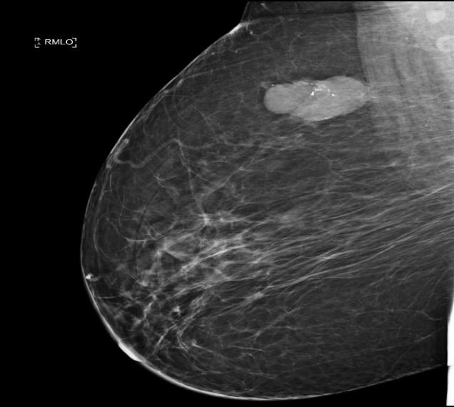
Right mammogram: Mixed density breast with a well-defined mass in the UOQ measuring 52 × 31 × 26 mm (Fig. 1).
-
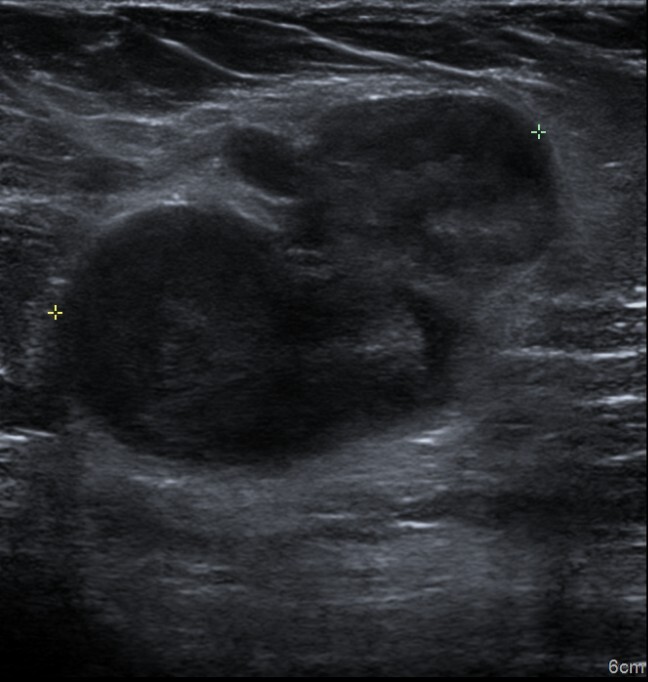
Right breast ultrasound: Large hypoechoic mass in the UOQ measuring 29 × 28 × 27 mm, showing a reduction in size from the previous examination (Fig. 2).
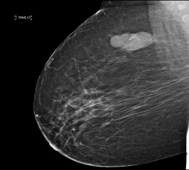
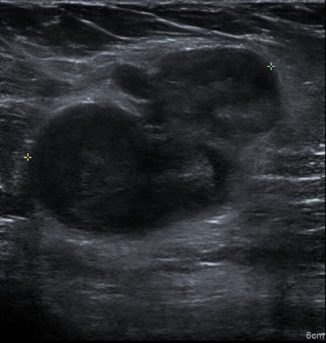
These follow-up findings suggested a poor response, with the persistent hypoechoic mass interpreted as likely residual tumour.
The case was discussed at the multidisciplinary team (MDT) meeting. As she was a PALB2-positive gene carrier, the patient underwent bilateral mastectomy and right sentinel lymph node biopsy. Gross examination of the right mastectomy specimen revealed a well-defined whitish indurated area measuring 45 × 40 × 25 mm. The left breast showed no significant abnormality.
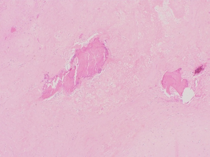
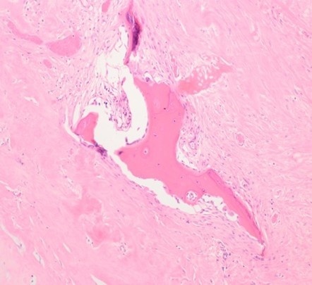
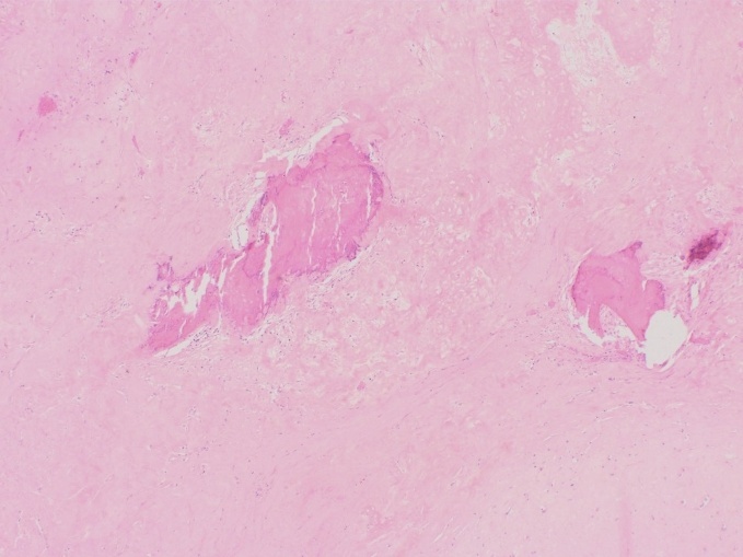
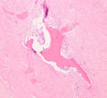
Histological sections of the right breast demonstrated widespread treatment-related changes, including diffuse fibroelastosis, chronic inflammation with foamy histiocytes, haemosiderin deposition, and foci of mature lamellar bone formation—consistent with osseous metaplasia (Fig. 3, 4). The remainder of the breast showed fibrocystic change. Importantly, no residual invasive or in situ carcinoma was identified in the breast or axillary lymph nodes.
3. Discussion
BOM is defined as extra-skeletal bone formation within soft tissue.1,2 This is an extremely rare entity and may pose a diagnostic challenge both clinically and radiologically.4 Our case represents an unusual occurrence of osseous metaplasia following neoadjuvant chemotherapy for breast carcinoma, without residual tumour. In this patient, post-treatment imaging interpreted the ossified focus as residual malignancy.
The presence of bone in post-treatment breast tissue is rare and may result from:
-
Chemotherapy-induced stromal changes and necrosis
-
Metaplastic transformation of stromal elements
-
Local activation of osteogenic pathways such as bone morphogenetic proteins (BMPs)
The pathogenesis of BOM is not fully understood. It has been postulated that osteoblasts may develop from modified stromal fibroblasts undergoing transformation under certain conditions.1,4
Literature review does not discuss BOM following neoadjuvant therapy for breast carcinoma.5–9 To our knowledge, no published case reports describe BOM arising specifically after chemotherapy in the absence of metaplastic carcinoma or other malignancies. Hyun Ho Lee et al. reported a case with lipogranuloma with benign osseous metaplasia in the breast following oriental medicine treatment for a benign lesion.10 Existing reports of BOM post-treatment typically involve malignant bone-forming tumours (e.g., matrix-producing carcinoma) rather than benign bone tissue as a reactive change. Benign bone-containing lesions are usually associated with benign tumours such as fibroadenoma and are not typically seen after chemotherapy.
This case illustrates the imaging challenge posed by benign metaplastic changes. Dense ossified areas can mimic tumour recurrence or persistence, particularly in the absence of pre-treatment correlation or biopsy confirmation. It underscores the importance of including benign post-treatment changes in the imaging differential diagnosis.
4. Conclusion
BOM of the breast is a rare but important diagnostic consideration following chemotherapy for breast cancer. This case demonstrates that such benign changes can closely mimic residual carcinoma radiologically and present a diagnostic challenge. A multidisciplinary approach with careful radiologic–pathologic correlation is essential in post-neoadjuvant settings to ensure accurate diagnosis and optimal patient management.
ETHICS STATEMENT
No patient consent was needed for publication as patient details were completely anonymised. The need for informed consent was waived due to the retrospective nature of the case report, and all data was de-identified. (neither the patient nor anyone else could identify the published information).
AVAILABILITY OF DATA AND MATERIAL
Data sharing is not applicable to this article as no new data were created or analyzed in this case report.
COMPETING INTERESTS
The authors declare that they have no known competing financial interests or personal relationships that could have appeared to influence the work reported in this paper.
FUNDING
No funding to declare.
AUTHORS CONTRIBUTIONS
NG drafted the manuscript, ST cross checked the case report, and JM collected the radiological findings.
ACKNOWLEDGEMENTS
The authors extend their gratitude to Dr Vivek Misra, Consultant Clinical Oncologist, The Christie Hospital, Manchester and Dr Chloe Wright, Consultant Chest Contouring and Oncoplastic Breast Surgeon, North Manchester General Hospital, Manchester Foundation Trust, UK for their assistance with oncological management and clinical review.