
1. Introduction
Amyand’s hernia is defined as the presence of the appendix within an inguinal hernial sac. It was first described in 1735 by the surgeon Claude Amyand.1 Its incidence is low, occurring in approximately 0.19% to 1.7% of all hernias, and it is associated with appendicitis in only 0.07% to 0.13% of cases.2 It is even rarer when associated with recurrent inguinal hernia. It is difficult to diagnose and, in most cases, is identified intraoperatively.3 A definitive approach to intervention has not yet been established.4
The Losanoff and Basson classification has been reviewed for this case.3
2. Case presentation
A 45-year-old male patient with a history of open right inguinal herniorrhaphy using the Rutkow-Robbins technique three years prior presented with a three-day history of right inguinoscrotal pain associated with a palpable mass. Physical examination revealed a heart rate of 78 beats per minute, respiratory rate of 16 breaths per minute, blood pressure of 117/70 mmHg, and a temperature of 36.2°C. No signs of peritoneal irritation were observed. Physical examination revealed a transverse scar in the right lower quadrant from the previous surgery and a tender, irreducible, erythematous right inguinoscrotal protrusion. Initial laboratory results showed a hemoglobin level of 14 g/dL, hematocrit of 41.2%, white blood cell count of 9.45 × 10⁹/L, platelet count of 256 × 109/L, glucose level of 101 mg/dL, creatinine level of 0.77 mg/dL, blood urea nitrogen level of 14.33 mg/dL, and urea level of 30.7 mg/dL.
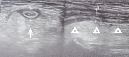
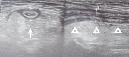
An extra-institutional ultrasound showed an incarcerated right inguinal hernia with the appendix within the hernial sac and associated inflammatory changes, leading to the diagnosis of Amyand’s hernia. (Figure 1).
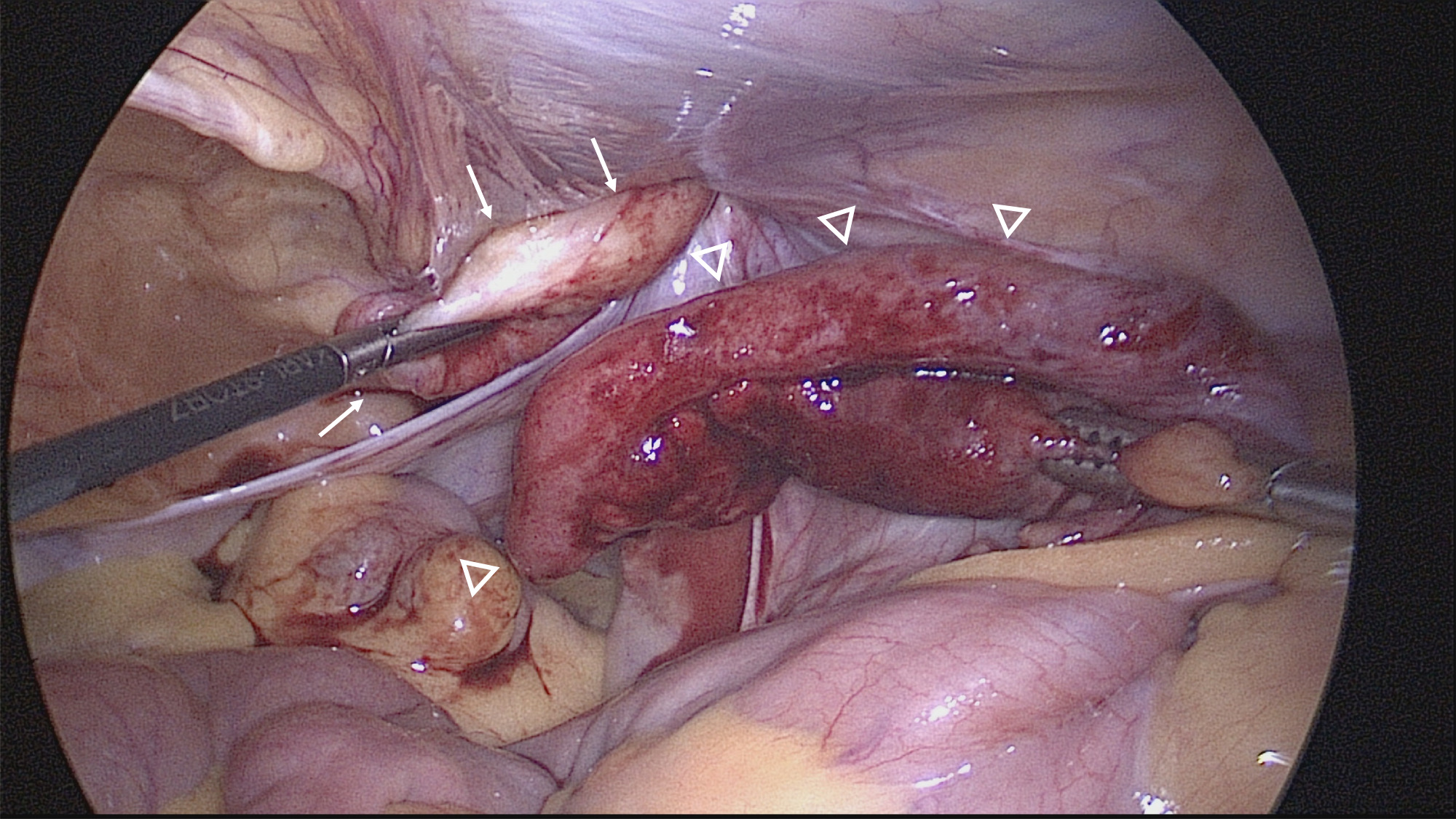
The patient is taken to surgery, prophylaxis is performed with Cefazolin 2 grams intravenously. A minimally invasive transabdominal preperitoneal (TAPP) approach was performed. A bloody fluid is found in the pelvis and an incarcerated indirect right inguinal hernia with the cecal appendix inside the hernial sac with inflammatory changes but without perforation, in addition there is bloody fluid in the hernial sac without purulent material (Figure 2).
_and_the_inflamed_cecal_.png)
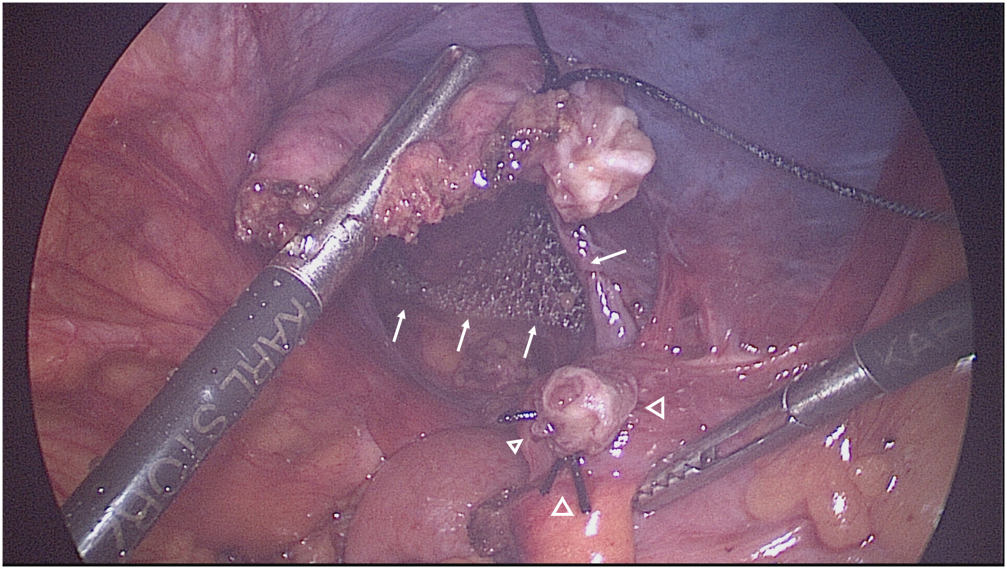
The patient received 3 g of ampicillin-sulbactam intraoperatively. The appendix was reduced from the hernia sac. Firm adhesions between the hernia sac and the previously implanted mesh were identified, requiring adhesiolysis and partial mesh explantation. The hernia was repaired using a transabdominal preperitoneal (TAPP) approach. An anatomically shaped large-pore polypropylene mesh (Bard 3DMax™ Extra Large, 12 × 17 cm; Warwick, RI, USA) was placed on the right side (Figure 2, Panel B) and secured with an absorbable fixation device. Subsequently, a laparoscopic appendectomy was performed. The appendiceal stump was secured using endoloops, and the appendix was retrieved in a specimen retrieval pouch. The operative time was 130 minutes, and the estimated blood loss was 20–30 mL.
_and_cecal_appendix_stump_with_endoloop_ligature_(arrowhead).png)
Postoperatively, the patient received intravenous ampicillin-sulbactam (3 g every 6 hours) for 72 hours. Laboratory tests performed on postoperative day 3 were within normal limits, and the patient was discharged without complications. Histopathological examination demonstrated acute appendicitis, characterized by fibrinopurulent exudate covering the appendix and abundant neutrophilic infiltration extending through the full thickness of the appendiceal wall to the serosa. No evidence of malignancy was identified.
The patient was evaluated in the outpatient clinic 10 days after surgery and demonstrated an uneventful recovery. At the five-month follow-up, there was no evidence of hernia recurrence, chronic inguinal pain, or mesh-related complications. No postoperative imaging studies were performed.
3. Discussion
The management of recurrent Amyand’s hernia associated with acute appendicitis using laparoscopic appendectomy and mesh repair is exceptionally rare. Consequently, the available evidence is limited to isolated case reports and small case series, precluding robust evidence-based recommendations.5,6
Amyand’s hernia associated with acute appendicitis and recurrent inguinal hernia represents a therapeutic challenge for the surgeon. This condition is usually diagnosed incidentally during surgery, with only approximately 17.2% of cases diagnosed preoperatively.2 Treatment requires appendectomy and hernia repair.
Because patients typically present with an incarcerated inguinal hernia, most surgeons proceed directly to surgery without obtaining additional imaging studies. However, in the acute setting, ultrasonography (47.6%) and computed tomography (70.4%) can identify the appendix within the inguinal canal,6,7 facilitate the differential diagnosis of the hernia sac contents, and detect associated complications, all of which are essential for planning the surgical approach. Regarding the diagnostic sensitivity of ultrasonography, we found no studies specifically evaluating Amyand’s hernia. However, ultrasonographic diagnosis is highly operator-dependent.
In our case, the patient was classified as having a type 2 Amyand’s hernia according to the Losanoff and Basson classification, a category traditionally associated with an increased risk of prosthetic mesh infection.6 It should be noted that when this classification was published in 2003, laparoscopic appendectomy and laparoscopic inguinal hernia repair were not yet widely adopted, and mesh repair was performed by only a minority of surgeons. Since then, numerous studies have demonstrated the advantages of the laparoscopic approach, including reduced postoperative pain, lower rates of surgical site infection, and faster recovery. Consequently, its use has increased substantially in recent years for both appendectomy and inguinal hernia repair, becoming a safe and effective option even in complex clinical scenarios such as the present case.
Given the recurrence following a previous anterior mesh repair and the presence of concomitant acute appendicitis, a transabdominal laparoscopic approach was selected to treat both conditions during the same procedure. This approach avoided re-entering the previous operative field, thereby minimizing dissection through scar tissue and adhesions while allowing placement of a new mesh in the preperitoneal space, thus avoiding direct contact between the mesh and the intra-abdominal viscera. Furthermore, in the absence of preoperative imaging, an exclusively totally extraperitoneal (TEP) approach would not have allowed exploration of the intraperitoneal cavity, potentially precluding the diagnosis of concomitant appendicitis.
In the presence of significant surgical field contamination, treatment options include primary tissue repair without mesh or the use of absorbable biologic mesh; however, both strategies are associated with higher rates of hernia recurrence. Notably, in the study by Manatakis et al., patients who underwent synthetic mesh repair did not develop surgical site infections or mesh-related complications, suggesting that synthetic mesh may be a safe option even in the setting of moderate contamination.
Transabdominal appendectomy and TEP hernioplasty have been described in a single surgical procedure, although few cases have been published. The extraperitoneal approach for hernia treatment has the advantage of using a different access route, reducing the possibility of prosthesis infection by using separate approaches for each procedure.
Current studies suggest that mesh repair is safe in contaminated surgical fields, even in the presence of perforated appendices and collections,3,4 without increasing infection rates, although this has been primarily described in incisional hernia repair. In cases of Amyand’s hernia, mesh repair appears to be a safe option in the absence of appendiceal perforation or purulent contamination; however, this approach has not been widely adopted, as most interventions are still performed using an open approach.4
The sectioning of the appendix, which represents the contaminated part of the surgery, was deferred until the end of the procedure.
Broad-spectrum beta-lactam antibiotics were administered for 72 hours because of the perceived risk of prosthetic mesh infection. This decision was based on the surgeon’s clinical judgment, acknowledging the lack of robust evidence supporting prolonged antibiotic therapy in this setting. Current evidence on antibiotic prophylaxis generally supports the administration of a single perioperative dose; however, some authors have advocated extended antibiotic therapy when synthetic mesh is used in the setting of acute appendicitis, suggesting that it may reduce postoperative complications.5 Our patient had an uneventful postoperative course without infectious or mesh-related complications.
In the present case, recurrent Amyand’s hernia was successfully managed with single-stage laparoscopic transabdominal preperitoneal (TAPP) repair with mesh and appendectomy. Nevertheless, management should be individualized,6,8 taking into account the degree of surgical field contamination, the severity of appendiceal inflammation, local operative findings, and the surgeon’s experience. In carefully selected patients, a single-stage laparoscopic approach may safely address both conditions while avoiding a second surgical procedure. To date, only a limited number of cases managed using this strategy have been reported.
Ethics approval
Written informed consent was obtained from the patient for the publication of this case report.
Availability of data and material
The data that support the findings of this study are available from the corresponding author upon reasonable request.
Competing interests
The authors declare that they have no known competing financial interests or personal relationships that could have appeared to influence the work reported in this paper.
Funding
This research received no specific grant from any funding agency in the public, commercial, or not-for-profit sectors.
Author’s contributions
Conceptualization: Jaime O Gutierrez-Sanchez (Lead). Supervision: Jaime O Gutierrez-Sanchez (Lead). Methodology: Jaime O Gutierrez-Sanchez (Lead). Writing – review & editing: Jaime O Gutierrez-Sanchez (Lead), Gabriela Gutierrez-Almanzar (Supporting). Investigation: Sarquid F Romero-Ayala (Lead), Karen Ortega-Dorado (Lead). Writing – original draft: Sarquid F Romero-Ayala (Lead). Project administration: Gabriela Gutierrez-Almanzar (Supporting). Resources: Karen Ortega-Dorado (Lead). Data curation: Karen Ortega-Dorado (Lead).
Acknowledgements
None.