
Introduction
Ascending cholangitis is a potentially life-threatening infection of the biliary tree that most commonly arises in the setting of biliary obstruction or altered anatomy. It typically involves biliary stasis with subsequent bacterial ascent from the duodenum, leading to intraductal infection, inflammation, and, in severe cases, systemic sepsis. While the majority of cases are caused by enteric organisms such as Escherichia coli, Klebsiella species, and Enterococcus, atypical pathogens are increasingly recognized, particularly in patients with surgically altered biliary anatomy.1 Recent studies have shown the overall incidence of postoperative cholangitis after biliary reconstruction range from 10-13%, with increasing incidence over time.2 Hepaticojejunostomy creation involves anastomosing the biliary tree to the jejunum, and predisposes patients to enteric reflux and colonization with a broader spectrum of organisms, requiring consideration of more rare pathogens.
Among these atypical pathogens, Yersinia enterocolitica is a rare but noteworthy cause. Yersinia enterocolitica is a gram-negative coccobacillus primarily associated with gastrointestinal disease, including enterocolitis, mesenteric adenitis, and pseudoappendicitis. It is most commonly transmitted via contaminated food products, particularly pork, with patients often presenting following a 1 to 6 day incubation period after exposure with acute gastroenteritis accompanied by fever, RLQ abdominal pain, and diarrhea lasting up to 3 weeks.3 However, clinical manifestations can range from self-limited diarrhea to invasive disease, especially in patients with altered gastrointestinal anatomy, immunocompromised hosts, or those with iron overload states, where the organism’s siderophilic properties enhance virulence. In the rare case of *Yersinia right upper quadrant abdominal pain, and potential jaundice, typically after an initial presentation of prodromal yersiniosis.
Postoperative Yersinia infections, while rare, have been reported in various contexts beyond cholangitis. These include prosthetic joint infections following total hip and knee replacements, autologous transfusion, and Yersinia-caused intussusception.4–7 The organism’s ability to survive at lower temperatures and evade host immune responses with a delayed onset of symptoms may contribute to its persistence in postoperative settings. However, its overall incidence in surgical infections remains low, and its identification often requires a high index of suspicion due to its atypical presentation and the need for specific culture conditions.
In this report, we review a rare, potential case of cholangitis possibly due to Yersinia Enterocolitica in a previously healthy 41-year-old Brazilian man following pancreaticoduodenectomy, illustrating the diagnostic and therapeutic challenges of this novel postoperative infection.
Case Presentation
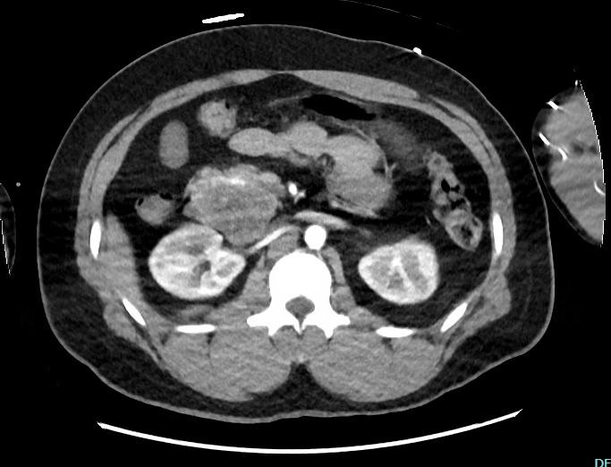
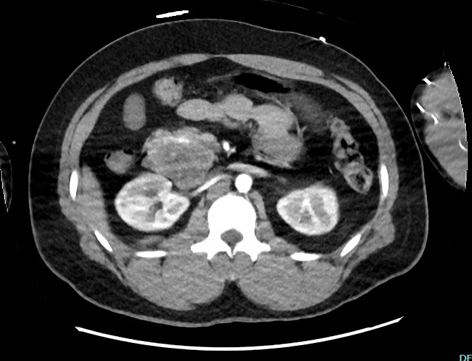
A previously healthy 41-year-old male who recently immigrated to the US from Brazil presented to the ED with hypotension and coffee ground emesis secondary to a 7 cm duodenal gastrointestinal stromal tumor Figure 1. CT angiogram of the abdomen and pelvis showed possible extravasation, and he was brought to the Interventional Radiology suite for mesenteric angiography and prophylactic embolization of his gastroduodenal artery.
Given the concern for recurrent bleeding, neoadjuvant imatinib targeting his Exon 11 C-KIT mutation was deferred, and he underwent upfront pancreaticoduodenectomy. Intraoperatively, he received Piperacillin-Tazobactam dosed every three hours. This patient’s duodenal GIST was antimesenteric and not obstructing the ampulla. He was noted to have a nondilated 3 mm hepatic duct. Estimated blood loss was 150 cc and no blood transfusion was given. A 19 French Blake drain was left posterior to the hepaticojejunostomy and pancreaticojejunostomy.
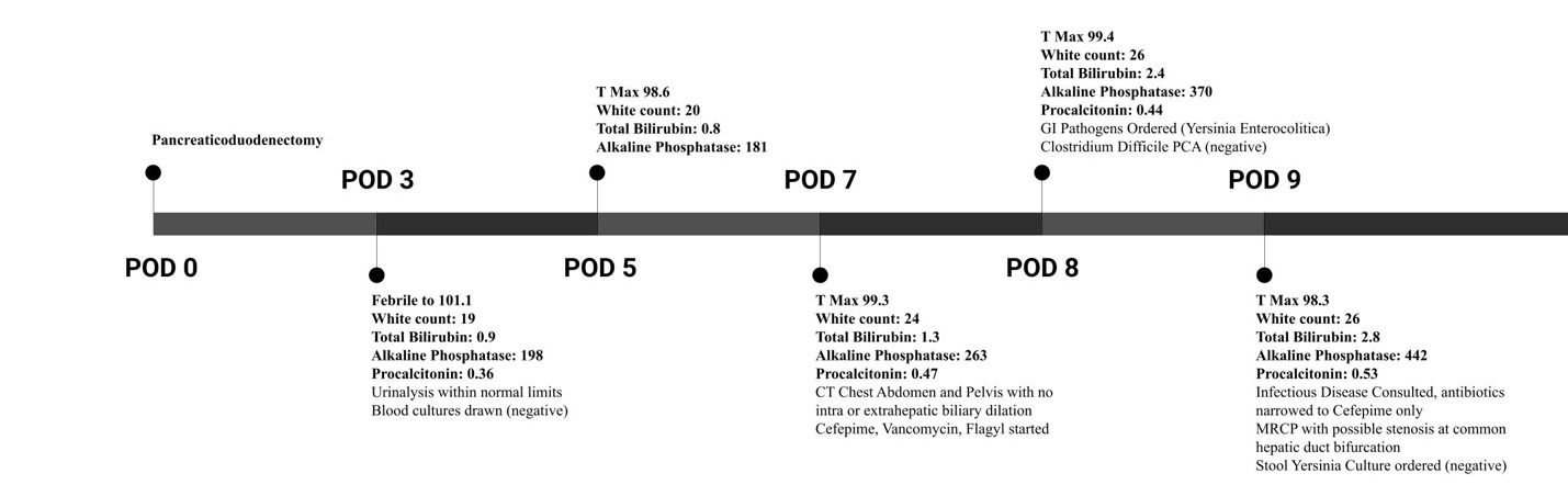
The postoperative period was complicated by a fever of 101.1 on postoperative day 3 and leukocytosis, up to 19 from 16. AST was mildly elevated at 77 and ALT was within normal limits. Vitals at this time included a heart rate of 126, blood pressure of 131/73, respiratory rate of 17 and oxygen saturation of 94%. His total bilirubin was 0.8 and alkaline phosphatase was 198. Laboratory values are trended in Figure 2.His urinalysis was within normal limits and lactic acid was normal. Blood cultures were drawn and resulted in no growth after 5 days. A Chest X-ray showed bibasilar opacities. Procalcitonin was 0.36. Drain amylase on POD 3 and 5 were 95 and 22 respectively, and the output remained nonbilious, sero-sanginous fluid.
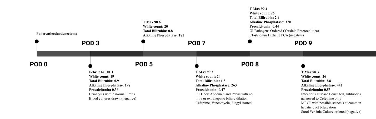
His leukocytosis continued to rise on POD 5 to 20, 22 on POD 6, 24 on POD 7, and 26 on POD 8. On POD 7, the patient began to report right upper quadrant discomfort with deep inspiration. He remained afebrile at this time, non-tachycardic with systolic blood pressure in the 120s. He also had multiple loose stools and 1 episode of emesis. Total bilirubin was 1.3 at this time and alkaline phosphatase was elevated at 263. CT Chest, Abdomen and Pelvis with PO and IV contrast was ordered to rule out pulmonary embolism and any intraabdominal pathology. Two 6 centimeter fluid collections were seen near the pancreaticojejunostomy. There was no intrahepatic ductal dilation. There were edematous changes in the pancreas consistent with pancreatitis, but his Lipase was 11. He was started on broad spectrum antibiotics with Cefepime, Vancomycin and Flagyl.
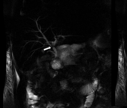
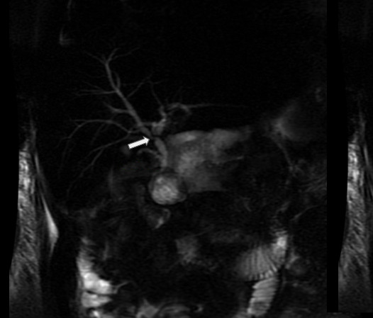
On POD 8, total bilirubin rose to 2.4 from 1.3, with a direct component of 2. Alkaline phosphatase also rose to 370. Clostridium Difficile PCR and stool GI pathogen PCR were ordered, which demonstrated Yersinia Enterocolitica. On POD 9, total bilirubin peaked at 2.8. Alkaline phosphatase was 442. Infectious Disease was consulted and an MRCP was ordered. The MRCP showed mild intrahepatic bile duct dilatation suggesting “stricture or stenosis at the bifurcation of the common hepatic duct into the right and left hepatic duct.” Stool cultures were ordered to corroborate the stool pathogens PCR results, however the Infectious Disease team also acknowledged a potential false negative result given the patient had been on antibiotic therapy for multiple days. The stool culture was ultimately negative. He was continued on Cefepime for Yersinia Enterocolitica coverage because of its known susceptibility to 3rd and 4th generation cephalosporins. His total bilirubin downtrended to 1 from 2.8 with conservative management. His white count also downtrended to 17 after peaking at 26. He was discharged home on Sulfamethoxazole/Trimethoprim, and scheduled for follow-up with Infectious Disease and Surgical Oncology. He has since recovered well with no recurrent episodes.
Discussion
Here, we present a unique case of post-pancreaticoduodenectomy cholangitis and gastroenteritis in a previously healthy 41-year-old Brazilian male who underwent pancreaticoduodenectomy for a duodenal gastrointestinal stromal tumor. Although postoperative cholangitis is a recognized early biliary complication, this case is noteworthy for the concurrent detection of Yersinia Enterocolitica by gastrointestinal pathogen PCR in his stool, raising the possibility of an atypical enteric pathogen contributing to an ascending infection in the setting of surgically altered biliary anatomy.
Common postoperative complications after pancreaticoduodenectomy include pancreatic fistula, delayed gastric emptying, and hemorrhage. Early biliary complications, including anastomotic leak or stenosis, cholangitis, and transient jaundice, occur in up to 14% of cases. Cholangitis is among the most frequent early biliary complications and typically manifests at least two weeks after surgery.8 Its pathophysiology in this setting often relates to anastomotic edema or stricture, afferent limb issues, ileus, or retrograde contamination of the biliary tree with enteric organisms in the absence of the sphincter of Oddi.
In this patient, the initial postoperative CT on POD 7 showed no intrahepatic ductal dilation. However, on POD 8, the total bilirubin rose from 1.3 to 2.4 mg/dL, prompting an MRCP. This demonstrated mild intrahepatic ductal dilatation, suggestive of possible anastomotic stricture or stenosis. Postoperative hyperbilirubinemia after pancreaticoduodenectomy can be multifactorial. Persistent elevation due to mechanical stricture generally requires intervention, whereas transient hyperbilirubinemia, as seen in this case, may result from anastomotic edema and often resolves spontaneously—as occurred here, without need for endoscopic or percutaneous intervention. An occult anastomotic leak was also possible, however no bile was ever seen in the drain, which was seated behind the hepaticojejunostomy.
Several factors in this case may have contributed to biliary complications. The patient had a small common bile duct (<5 mm) and no preoperative biliary obstruction or stenting.9 Preoperative chemoradiation, when applicable, and small-duct anatomy are recognized risk factors for postoperative biliary issues. Conversely, preoperative obstructive jaundice and biliary drainage are associated with lower rates of postoperative cholangitis, likely due to chronic proximal ductal dilation and wall thickening.8 Intraoperative common bile duct cultures were not obtained, per institutional protocol and published guidelines, in the absence of a preoperative stent.10 Routine CBD sampling may be worth considering in patients with small ducts or other known risk factors for biliary complications even without prior instrumentation.
While enteric gram-negative organisms account for most post-pancreaticoduodenectomy cholangitis cases, atypical pathogens are increasingly reported in patients with altered anatomy. In this patient, stool gastrointestinal pathogen PCR was positive for Yersinia Enterocolitica. His symptoms began on postoperative day 3, which follows the typical 1 to 6 day incubation period after exposure. Subsequent stool culture was negative after several days of broad-spectrum antibiotics. In addition to his altered biliary anatomy, this patient also received intravenous iron supplementation postoperatively, another known risk factor for enhanced virulence of Yersinina Enterocolitica, and was in a relatively immunocompromised state during the early postoperative period.
Importantly, we cannot definitively attribute the cholangitis or hyperbilirubinemia to Yersinia Enterocolitica, as common bile duct bile cultures—the gold standard—were not obtained. Gastrointestinal pathogen PCR panels have not been formally validated against bile cultures in cholangitis and should be interpreted with caution. Nevertheless, in patients with surgically altered biliary drainage, a positive stool PCR may help identify dominant enteric pathogens and inform antibiotic selection when bile cultures are not available. Emerging data also suggests that the fecal microbiome may influence postoperative outcomes after hepatopancreatobiliary surgery.11
The patient was transitioned to Trimethoprim-Sulfamethoxazole, an oral agent to which Yersinina enterocolitica is usually susceptible, after clinical improvement with normalization of his bilirubin and resolution of his loose stools and abdominal discomfort.12 He has since recovered as an outpatient and has initiated consultation for adjuvant imatinib therapy targeting his exon 11 C-KIT mutation, given the elevated risk for recurrence associated with small bowel location.
This case highlights the diagnostic challenges of postoperative hyperbilirubinemia and the potential role of molecular stool testing in guiding management when atypical pathogens are suspected, while underscoring the limitations of attributing causality without direct biliary cultures.
Consent
Written informed consent was obtained from the patient for publication of this case report and accompanying images.
Availability of data and material
The data that support the findings of this study are available from the corresponding author upon reasonable request.
Competing interests
The authors declare that they have no known competing financial interests or personal relationships that could have appeared to influence the work reported in this paper.
Funding
This research received no specific grant from any funding agency in the public, commercial, or not-for-profit sectors.
Authors’ contributions
AH, NM, and RZ contributed equally in drafting the manuscript. BG and AR contributed equally in editing the manuscript. EP drafted and edited the manuscript.