
1. Introduction
Upper gastrointestinal bleeding (UGIB) is a life-threatening pathology that involves bleeding proximal to the ligament of Treitz. This pathology is relatively common and is associated with high mortality in the ED. In fact, UGIB has been estimated to account for up to 20,000 deaths annually in the United States.1 While some patients present with frank hematemesis, many present with more subtle symptoms such as melena, vague abdominal discomfort, nausea, or symptoms of anemia, and can deteriorate quickly into hemorrhagic shock. Because early clinical findings may be nonspecific, patients with UGIB require expeditious diagnosis and early resuscitation in the ED. Gastric point-of-care ultrasound (POCUS) can support early diagnosis and risk stratification to prioritize monitoring, expedite transfusion, and facilitate urgent endoscopic intervention.
Gastric POCUS is an underutilized bedside technique in the assessment of UGIB in the ED. Although POCUS has an established role in cardiovascular emergencies, aortic pathology, respiratory failure, trauma resuscitation, and vascular access, the role of this bedside imaging modality in UGIB and aspiration risk assessment in the ED remains relatively uncommon and less well-studied.2–6 Emerging literature suggests that gastric POCUS can provide immediate supportive evidence to identify intragastric blood, detect patients requiring immediate escalation of care, and assess for aspiration risk when paired with appropriate clinical assessment in the ED.7–9 Here we present a case of insidious UGIB in which gastric POCUS enabled rapid diagnosis, early triage, and stabilization.
2. Case Presentation
A 58-year-old male with a past medical history of hypertension and type 2 non-insulin-dependent diabetes presented to the ED with 2-3 days of diffuse abdominal pain associated with nausea and melena. The patient had not eaten since the night before. On arrival, the patient was ill-appearing with a blood pressure of 95/64 mmHg, heart rate of 75 bpm, respiratory rate of 16, and oxygen saturation of 98% on room air. Physical exam was notable for diffuse abdominal tenderness without peritoneal signs.
__and_sagittal_views_(b)_visualiz.png)
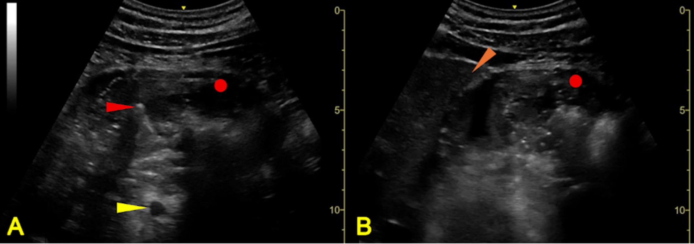
Initial gastric POCUS demonstrated findings concerning for intragastric blood, prompting immediate escalation of care (Figure 1). The patient was transferred from the triage to critical care zone with airway precautions including suction at bedside, head-of-bed elevation, and intubation equipment. Several minutes after transfer of the patient to the critical care zone, the patient subsequently developed an episode of hematemesis with suction at bedside and head-of-bed elevation helping to maintain airway protection. Laboratory findings subsequently revealed normocytic anemia with hemoglobin 7.5 g/dL, leukocytosis of 17.5×10³/µL, BUN 41 mg/dL, creatinine 1.0 mg/dL, and elevated lactate of 4.6 mmol/L, consistent with hemorrhagic shock secondary to UGIB on gastric POCUS.
Gastroenterology was immediately consulted after sonographic assessment, and the patient was treated with 2 units of packed red blood cell transfusion, intravenous pantoprazole, octreotide infusion, and ceftriaxone. The patient was admitted to the intensive care unit for close monitoring and subsequent endoscopy identified a gastric ulcer with a visible vessel (Forrest IIa) that was successfully treated with endoscopic clipping. The patient recovered without further complications.
3. Discussion
In this case, gastric POCUS was used to rapidly diagnose an insidious UGIB with hemorrhagic shock. Identification of intragastric blood on gastric POCUS altered management of the patient by facilitating immediate escalation of care, including initiation of aspiration precautions with transfer of patient to a critical care zone, preparation for blood transfusion, earlier administration of medical therapy, and rapid consultation with gastroenterology for definitive endoscopic management. Emerging literature suggests that gastric POCUS can provide clinically relevant information regarding gastric contents and aspiration risk and may serve as a useful tool for risk stratification and management in the ED.7–9 In prior reports, gastric POCUS identified gastric blood in patients presenting with hypotension with subsequent confirmation via enteric tube aspirate and assessed for aspiration risk after confirming UGIB in patients with reported hematemesis or evidence of UGIB on clinical exam.7,8 Here, gastric POCUS identified gastric blood in an initially stable patient in a fasted state with a nonspecific presentation, rapidly escalating care and implementing aspiration precautions prior to hematemesis.
Gastric POCUS is performed by first scanning the gastric antrum in the supine position and then in the right lateral decubitus (RLD) position.7–9 The RLD position allows for the most sensitive and specific evaluation of gastric volume by shifting gastric contents into the antrum, which is the most gravitationally dependent portion of the stomach (Figure 1).8 This produces the most reliable location for qualitative and, if needed, quantitative, assessment of volume and contents of the stomach. Although the RLD position may not always be feasible, particularly in critically ill patients, a full and distended stomach in the supine position may be sufficient to rule in a distended antrum (Figure 1). Generally, the low-frequency (2–5 MHz) curvilinear transducer probe is utilized due to the wider field of view and deeper penetration depth associated with this probe.8,9 The transducer is placed in the subxiphoid area or epigastrium in a sagittal orientation with the probe marker pointed cephalad and then swept from the left to the right subcostal margins to obtain sonographic views of the stomach.
Various contents of the stomach have different sonographic characteristics. For example, physiologic gastric secretions and clear fluids, such as water and black coffee, appear hypoechoic or anechoic and can be associated with co-ingested gas bubbles that appear as multiple free-moving, minute echoes with a “starry night appearance.”7–9 In comparison, thicker fluids, such as milk, appear with higher echogenicity.9 In contrast, the initial appearance of recently ingested solid meal can appear with a frosted-glass pattern, which is visualized as a mucosal-air interface along the anterior stomach wall, associated with a “ring-down” artifact that blurs deeper contents and the posterior wall of the stomach.9 As food is digested, this interface dissipates as air is displaced and the stomach contents become heterogeneous with mixed echogenicity.9 Importantly, this heterogeneous appearance mirrors the sonographic appearance of blood clots in UGIB, where blood clots may appear as irregular hyperechoic densities free-floating within a distended lumen of anechoic nonclotted blood, with dynamic movement of gastric content representing active extravasation (Figure 1).7,8 Because sonographic findings suggestive of UGIB mirror the appearance of stomach contents in a patient who has recently eaten food or drank a beverage, appropriate clinical context is essential in interpreting sonographic findings.
While the role of gastric ultrasound in aspiration risk assessment has been well-investigated in the field of anesthesiology, studies evaluating its role in the ED remain limited.7–12 There is also a paucity of data directly linking sonographic gastric findings or estimated gastric volume to aspiration events in patients with UGIB.8,9,13–15 In the emergency setting, a qualitative assessment of gastric volume is more expedient and feasible to quickly provide information that can influence ED decision making and aspiration risk. For example, sonographically visualizing a full, distended stomach or one that contains solids, such as food or blood clots, may suggest that the patient may be at high risk for aspiration without the need for quantitative assessment (Figure 1).8,9 Conversely, an empty gastric antrum in the RLD position, visualized sonographically as a “target-shaped” small hyperechoic oval shape with a collapsed, thick, multilayered, hypoechoic wall, suggests low aspiration risk.8,9 When neither of the above extremes are visualized, the provider can repeat imaging to reassess for a qualitative increase in gastric antrum size, which may suggest ongoing bleeding. This allows for assessment of real-time changes other imaging modalities do not allow. Additionally, the provider can perform quantitative measurements to assess for aspiration risk.8,9 Although the exact gastric volume associated with increased aspiration risk remains contentious, clinical data in the setting of anesthesia proposes that a gastric volume greater than 1.5 ml/kg is commonly used as a threshold to define a high-risk stomach for aspiration, and several mathematical equations have been employed in calculating this volume.8,9,15
Identification of a patient with an UGIB and high aspiration risk may help individualize decisions regarding airway management. For example, a patient with intragastric blood on POCUS may prompt head-of-bed elevation, suction readiness, nasogastric tube placement, and preparation for potential intubation and nasogastric tube placement. Importantly, while identification of a distended stomach may correlate with high aspiration risk, this does not necessitate endotracheal intubation. Rather, gastric POCUS allows for earlier airway preparation and closer monitoring. If a patient is not protecting their airway, gastric POCUS can prompt consideration for varied airway techniques, such as Suction Assisted Laryngoscopy and Airway Decontamination and Head-Elevated Intubation.16,17 While both an empty stomach and a patient with preserved mental status and an intact airway despite a full stomach may support the decision to defer intubation, the decision to perform prophylactic intubation in patients with UGIB and elevated aspiration risk remains controversial. For example, some studies suggest that routine intubation before endoscopy may be associated with higher rates of pneumonia, longer hospital stays, and worse outcomes.18
Although gastric POCUS can support clinical decision making, utilization of this imaging modality requires familiarity with characteristic sonographic imaging findings in conjunction with operator experience and expertise.8 Additionally, a fasting patient is critical for accurate interpretation.7–9 Furthermore, while gastric POCUS has potential in rapid diagnosis and assessment of aspiration risk, this imaging modality cannot distinguish between different sources of bleeding, such as esophageal varices or gastric ulcers. As case reports are inherently anecdotal in nature, future research is necessary to validate the potential of gastric POCUS in assessment of aspiration risk and stabilization of patients with suspected UGIB. Prospective studies on the impact of gastric POCUS on time to endoscopy, as well as studies investigating the interobserver reliability of sonographic imaging findings are essential in validating the potential utility of gastric POCUS.
4. Conclusion
Gastric POCUS is an underutilized bedside tool that may support early diagnosis, triage, and management of suspected UGIB in the ED. In the appropriate clinical context of a fasting patient with suspicion for UGIB, gastric POCUS can rapidly identify intragastric blood, facilitating aspiration precautions with airway planning as well as ensuring timely resuscitation and specialist consultation. Furthermore, this case demonstrates the potential use of gastric POCUS as a screening tool for UGIB in an undifferentiated hypotensive patient when UGIB is suspected based on clinical findings of melena, hematemesis, and presence of risk factors such as alcoholism and hepatic disease.7,8 Although the utility of gastric POCUS may be constrained in cases of limited history and occult UGIB, when incorporated into screening protocols such as the Rapid Ultrasound for Shock and Hypotension (RUSH) examination, gastric POCUS can potentially facilitate earlier recognition of UGIB and influence management decisions in the appropriate clinical context.
Declarations
Ethics approval
Written informed consent was obtained from the patient for publication of this case report and accompanying images. This case report was reviewed and approved by the institutional ethics committee.
Availability of data and material
Data sharing is not applicable to this article as no new data were created or analyzed in this study.
Competing interests
The authors declare that they have no known competing financial interests or personal relationships that could have appeared to influence the work reported in this paper.
Funding
This research received no specific grant from any funding agency in the public, commercial, or not-for-profit sectors
Authors’ contributions
DH conceived the study. TR, BK, and DH drafted the manuscript. All authors contributed substantially to its revision.
Acknowledgements
Disclaimer: This research was supported (in whole or in part) by HCA Healthcare and/or an HCA Healthcare affiliated entity. The views expressed in this publication represent those of the author(s) and do not necessarily represent the official views of HCA Healthcare or any of its affiliated entities.