
Introduction and Background
The story of the fMRI’s creation is tied inextricably to the story of our developing understanding of the brain as a whole; as fundamental to our knowledge of the brain as the ruler is to geometry. Previously, brain activity was foreign territory, as tracking its activity in a conscious human was completely impossible. Given how fundamental it is as a tool of observation and measure, its existence will likely enable research just as groundbreaking as the findings that shaped it. Because of its current and future importance, understanding the history and inner workings of the fMRI is crucial.
Review
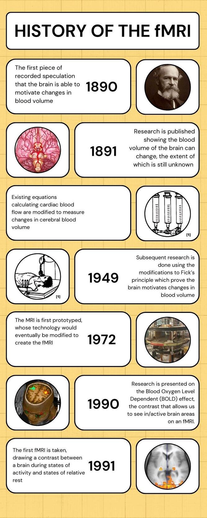
The basic underpinning behind the fMRI, the idea of blood flow predicting brain activity, is much older than the technology itself. The first recorded mention of this idea comes in William James’ 1890 work, The Principles of Psychology, where he mentions an earlier experiment conducted by an Italian colleague. By placing a person on a scale and eliciting intellectual activity, the researcher found subtle changes in weight towards the head-end of the scale and concluded that intellectual activity redistributed blood toward the brain.2 While conventional wisdom will tell us that there was likely very little change in weight if any at all, the fact remains that this idea has been circulating the collective scientific consciousness since at least then.
To that point, although a minority of scientists believed that the brain controlled the flow of blood through its activity level, the prevailing belief at the time was that the rigidness of the skull made changes in the amount of blood in the brain impossible entirely. In an effort to prove the contrary, two British researchers used a purpose-made device to measure the changes in cerebral blood volume among anesthetized dogs.3 While their findings succeeded in showing that there were meaningful changes in cerebral blood volume, their results were mostly qualitative rather than quantitative, meaning there was no way to measure the extent to which this was the case. They also failed to prove that the brain itself played a role in driving this change in blood volume. Although the two researchers tried to link the changes to chemical impulses, their lack of quantitative data made proving that link impossible.4
Research on the topic of tracking blood flow to the brain would continue, and although consequent findings further disproved the idea that the skull stops blood from flowing into and out of the brain, nobody had yet been able to prove the fact that cerebral brain flow changed as a result of brain activity. To that point, nobody had been able to quantitatively measure cerebral blood flow in a conscious person under normal bodily conditions. As a result, the conventional belief remained that external factors governed blood flow to the brain, rather than the brain itself, and it remained that way for over 50 years. The first concrete piece of evidence supporting the idea that the brain was the regulator of blood flow came as probably the most important piece of research in the pathway to the creation of the fMRI. In a 1948 publication, researchers devised a novel technique to quantify cerebral blood flow noninvasively in a conscious human. Their methodology was based on Fick’s principle, which states that cardiac output is equal to the consumption of pure gaseous oxygen (in milliliters) per minute divided by the oxygen content of arterial blood minus the oxygen content of mixed venous blood final equation: 5 Put most simply, Fick’s principle is used to quantify the amount of blood flowing into an organ. The biggest issue with Fick’s principle when attempting to create novel calculations is that it contains three variables, making it hard to isolate any two and do algebra. The researchers realized that they could eliminate the consumption of gaseous oxygen by using a blood constituent the body couldn’t rapidly metabolize, as opposed to how the body interacts with oxygen. They settled on nitrogen as an alternative, and by measuring the arterial and venous concentrations of nitrous oxide rather than oxygen, the two were able to jump the largest hurdle thus far in settling the debate over the brain’s ability to intrinsically motivate cerebral blood flow: a reliable, quantitative measure of cerebral blood flow in a person under normal conditions.6 In subsequent papers, not only were the two able to use their new method of measure to establish a link between inhaling oxygen and changes in cerebral blood flow, but they were also able to prove that these changes were motivated by chemical signals released as a result of increases or decreases in neuronal oxygen use.7
Any discussion of the function of modern fMRI technology is incomplete without at least a rudimentary understanding of its more versatile, less specialized older sibling, Magnetic Resonance Imaging (MRI). MRIs are large cylindrical machines that play host to a complex system of magnets at differing levels of strength used to create detailed maps of the soft tissues in the body. Every water molecule releases tiny electromagnetic pulses constantly, and as it turns out, there are quite a few water molecules in the human body. MRI machines create a large magnetic field surrounding the body of the person being scanned, which uses the electromagnetic pulses in those water molecules to orient their protons in the same direction.8 Once those molecules are all oriented the same, the MRI machine will periodically pulse out a weaker electromagnetic field encouraging them to point in a different direction, causing a tiny change in their orientations. In effect, the weaker magnetic field causes these molecules to slightly twitch in place. This slight twitch releases recordable radio-level photons, the intensity and quickness with which they are recorded varying highly with the strength of the magnetic impulse and thus the thickness of the tissue. These differences in depth allow us to create the detailed, three-dimensional images MRI machines are valued so highly for.9 The fMRI modifies this function by changing how the images are interpreted. As evidenced by the work done using Fick’s principle, brain activity is most easily predicted by the flow of oxygen-rich blood to the areas being engaged. As it turns out, the electrons in oxygenated blood tend to block out the magnetic fields engaged by MRIs, stopping the protons in the hydrogen molecules from quickly aligning in the encouraged direction. Deoxygenated blood does not behave similarly, meaning more photons are released when the machine is reading areas that are deoxygenated compared to areas that are oxygenated.10
The specifics of the development of the MRI deserve their own paper entirely, but the machine we have come to know was first prototyped in 1972.11 The most foundational piece of research detailing the modifications made to the interpretations of an MRI when conducting an fMRI didn’t come until the early 1990s when new research was presented on the blood oxygenated level dependant (BOLD) effect.12 The paper was the first to describe the contrast in imagery between areas of the brain flooded with oxygenated blood and deoxygenated blood, allowing for the levels of oxygen throughout the brain to be measured in real-time, creating a map of neuronal activity. The first fMRI conducted during a state of intellectual stimulation was taken in 1991 when researchers could use existing MRI technology to map cerebral blood volume during both rested and stimulated cognitive states.13
Since that landmark work, fMRIs have been crucial in expanding our understanding of how the brain works: as of the time of writing, 786,406 entries on PubMed mention “fMRI”, and many of the most critically important new works in furthering our knowledge of the brain use the technique as a key method of gathering data. In clinical settings, fMRIs are used to plan brain surgeries, which are much safer and more successful now that they are conducted with detailed views of brain activity to reference, as well as to inform treatment plans.14
As important a tool as the fMRI is, our understanding of its usage remains fairly rudimentary compared to its potential. Researchers continue to search for more ways to interpret the data an fMRI presents: whether that means learning more about cognitive state, markers belying disease, or monitoring neurological disorders. Importantly, as fMRIs continue to grow cheaper and more accessible, care will become more personalized, as the ability to map out brain activity in response to certain stimuli allows for the customization of pain management techniques and the management of mood disorders, among other applications.15
Conclusion
Over the course of 100 years, neurologists went from speculating on the brain’s ability to modulate its blood flow to using the nature of that flow to map out the brain’s activity. We’ve been using and iterating on the technology extensively in the 35 years since, and the scope of its potential uses still dwarfs what we can currently do. The fMRI is the culmination of a generation of learning on our understanding of the brain, and the nature of the technology means that it has the potential to drive similarly groundbreaking work in the future.